-
E-mail
weiscope@163.com
-
Phone
13538978162
-
Address
802, 8th Floor, Quanfeng Commercial Building, No. 8 Erheng Road, Yuancun, Tianhe District, Guangzhou City
Product Categories
Guangzhou Weiyu Optical Instrument Co., Ltd

Microscopic detection of fish diseases
NegotiableUpdate on 02/15
- Model
- Nature of the Manufacturer
- Producers
- Product Category
- Place of Origin
Overview
Researchers are dissecting fish bodies for slicing. With the increasing scale of aquaculture, farmers are investing more and more, and the risks are also increasing. Therefore, scientific farming methods are essential
Product Details
Researchers dissect fish bodies to make slices
With the increasing scale of aquaculture, farmers are investing more and more, and the risks are also growing. Therefore, scientific breeding methods are essential. For fish farming professionals, once fish become ill, there will be a widespread infectious effect, ultimately leading to widespread illness or death, and the losses to the farmers will be enormous. To control fish diseases, early prevention is necessary. In addition to visual observation, a more effective method is to use microscopic examination to determine what bacteria or viruses fish are infected with and prescribe the right medicine to prevent them in advance.
Inspection Method
1Observe the surface of diseased fish. Carefully inspect the mouth, eyes, scales, fins, and other parts of the diseased fish in order, and some obvious pathogens such as water mold, nematodes, fish carp, and spearhead fleas can be seen with the naked eye. Observe whether the mouth and surface of the diseased fish are bleeding, whether there is an increase in mucus on the surface, whether the anus is swollen, and whether the scales have fallen off or become erect. Check whether the fins of the diseased fish are intact, whether there are pus or flocculent attachments in the affected area (if necessary, scrape them off for examination), whether the eyes are protruding, and whether the eyes are cloudy.
2Gill examination. Firstly, place the fish naturally flat and check if the gill cover is open. Then, open the gill cover to check if there is any increase in mucus, congestion, decay, or mold on the gill pieces. Finally, use scissors to cut open the gill cover and remove a portion of the gill filaments for observation under a microscope.
3Anatomical examination. To examine the body of a fish, the first step is to dissect the diseased fish (as shown in the picture), use scissors to cut along the surface of the body from the anus, open the abdominal cavity, remove all internal organs, separate each organ, and then examine them one by one. Observe the internal organs for abnormalities, foreign objects or parasites, and whether there is ascites; Cut open the intestines and check for redness, swelling, and the presence of feed in the intestines
Live insects, etc; Observe whether each organ is normal, whether there is bleeding, discoloration, hardening, hypertrophy or atrophy.
4Microscopic examination. Based on the examination of diseased fish, sampling from key areas allows for intuitive and rapid observation of diseased organs, parasites, etc., with the characteristics of high diagnostic rate, fast and convenient. Generally speaking, the areas that require microscopic examination include surface mucus, gills, and intestinal mucus. Some parasites may be observed on the surface mucus, such as wheel worms, ciliates, small melon worms, fish bean worms, etc; Gills may show redness and swelling of gill filaments, increased mucus, and parasitic infections. There are many common parasitic infections in the gills, such as wheel worms, ring worms, ciliates, small melon worms, third-generation worms, and oblique tube worms; There are tapeworms, echinoderms, and myxosporidium that can be observed in the intestine. During inspection, the sample should be kept fresh and easy to observe. An appropriate amount of physiological saline can be dripped in, and the examination should be focused and rapid
The longer the interval, the harder it is to find the pathogen.
(1) Mucus: Examination of pathogens that are not visible to the naked eye in the mucus on the surface of fish, such as tremulous flagellates, oral worms, wheel worms, myxosporidium, and small melon worms, as well as fluke cysts.
(2) Gills: Check for the invasion and parasitism of various pathogens, bacteria, or parasites. Protozoa such as rotten gills, gill molds, gill flagellates, myxosporidium, microsporidia, dermatosporidium, wheel worms, oblique tube worms, small melon worms, half browed worms, tongue cup worms, and hairy tube worms, as well as single flukes such as ring worms, third generation worms, and double bodied worms, as well as metacercariae of compound flukes, larvae of mollusks, and carp. exist
(3) Gastrointestinal examination: Check the gastrointestinal tract for parasites such as flagellates, amoebas, myxosporidium, microsporidia, coccidiosis and other protozoa, as well as trematodes, nematodes, echinoderms, tapeworms, trematodes, tapeworms, nematodes and echinoderms, hexaflagellates, amoebas, and intestinal pocket worms
(4) Liver examination: Small white spots formed by the cysts or bodies of Paragonimus, Myxosporidium, Microsporidia, or Coccidia in the liver


The method of microscopic examination is to take a small amount of tissue or mucus from the lesion site and place it on a glass slide. If it is tissue or mucus from the body surface and gills, a small amount of ordinary water should be added; If it is visceral tissue, physiological saline is used, then covered with a cover glass and slightly flattened, and observed under a microscope (usually 2-3 glass slides need to be made). First, low magnification is used for examination, and when the structure of the insect body cannot be seen, medium or high magnification is used for examination. Slide from left to right, move a small grid, then from right to left, repeat the process from top to bottom until the entire slide is viewed, and record the types and quantities of parasites. A small amount of parasites does not cause fish diseases, but if three fields are randomly observed and counted on a glass slide, with an average of about 20 fields per field, attention should be paid. Parasites with a size of several tens of micrometers (such as flagellates) are usually counted at high magnification, while parasites with a size of several hundred micrometers (such as rotifers) are counted at low magnification.
Both visual and microscopic examinations should be conducted to assess the condition of the diseased fishcomprehensive analysisSome conditions are not caused by a single disease and should be analyzed and judged based on multiple factors.
According to the statistics of products used by a large number of fish farmers by Weiyu Company, the most suitable microscope for observing fish diseases should not only be able to clearly see the presence of parasites and bacteria in fish diseases, but also take into account the economic affordability of farmers. Products that are cheap but of lower quality cannot meet the application effect, while high-quality products are more expensive. The optimal magnification for fish disease observation is around 100x and 400x. Non researchers should not blindly pursue high magnification.
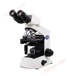
WSB300 Biological MicroscopeAs a cost-effective product that meets the above characteristics, it can be expanded into a digital microscope as needed and can save fish disease data for customers. It is most suitable for being recommended for fish disease detection and has served hundreds of professional breeders in the Yangtze River Delta, Pearl River Delta and other areas, receiving high praise from many customers.