-
E-mail
weiscope@163.com
-
Phone
13538978162
-
Address
802, 8th Floor, Quanfeng Commercial Building, No. 8 Erheng Road, Yuancun, Tianhe District, Guangzhou City
Product Categories
Guangzhou Weiyu Optical Instrument Co., Ltd

Traditional Chinese Medicine Biological Polarized Microscope WSB1200P
NegotiableUpdate on 02/15
- Model
- Nature of the Manufacturer
- Producers
- Product Category
- Place of Origin
Overview
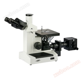
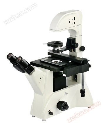
The WSB1200P biological polarizing microscope is a newly launched high-level microscope designed specifically for the detection of Chinese and Western medicinal materials in China. It has outstanding optical performance and multiple observation functions. It adopts the most advanced optical design, infinite distance optical system, semi apochromatic flat field infinite distance objective lens, and a large and flat field of view, thus obtaining high-quality optical imaging quality
Product Details
WSB1200P biological polarizing microscope specially designed for the detection of Chinese and Western medicinal materials
The newly launched high-level microscope in China has outstanding optical performance and multiple observation functions, adopting the most advanced optical design, Infinite distance optical system, semi apochromatic flat field infinite distance objective lens, with a large and flat field of view, resulting in high-quality optical imaging. It can be widely used in research institutes such as biology, bacteriology, histology, and medicinal chemistry, and is suitable for inspection in medical and health institutions, laboratories, research institutes, and higher education institutions.
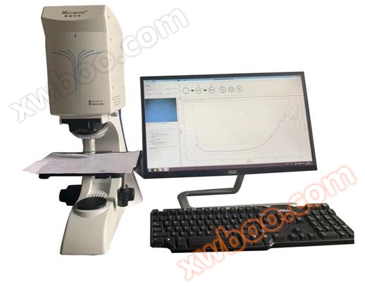
The system is equipped with a high-definition image sensing photography system, which processes, edits, saves, and outputs images through a computer, The system can further integrate with the microscopic image analysis system(WIMAGE X64 5.0Software),Perform metrological testing, morphological analysis, research statistics, and output graphic and textual reports on microscopic images. Covering almost all application areas of image quantitative analysis, various detection and analysis related to image morphology can be completed.
Technical parameters of biological microscope:
1. Optical system:CCISInfinite chromatic aberration independent correction optical system
2. Body: Integrated design, integral die-casting, more stable and sturdy body
3. Magnification factor:40~l000X
4. Eyepiece: with anti mold function, flat field10XHigh eye point eyepiece, high eye point observation.
5. Objective lens: Infinite achromatic objective lens (with anti mold function),4X/0.10;10X/0.25;40X/0.65 (with spring and buffer device);100X/1.25(Oil, equipped with springs and buffering devices).
6. Twisted binocular/Three eyes: infinite distance, observation angle30°, distance between the two pupils52mm~75 mmThe visual acuity is adjustable.
7. Objective lens converter: internal positioning four hole conversion structure;
8. Coarse and micro focusing device: Low position coarse motion coaxial focusing handwheel; Micro motion handwheel0.1mm/Conversion, grid value0.001mm;The coarse elasticity can be adjusted,14mm/Turn; Workbench upper limit device, maximum travel20mm;
9. Carrier platform:210mm×140mmEquipped with a movable ruler and a range of movement76×50mm, accuracy0.1mm;X、YAdjust the handwheel towards the lower coaxial position;
10. Spotlight: Abbe spotlightN.A.1.25Equipped with a phase contrast socket, a handwheel lift type, and a precise up and down adjustable system for the spotlight, the spotlight can be accurately matched with various magnifications of objective lenses. The spotlight bracket is equipped with a spotlight center adjustment device, which facilitates the alignment of the lighting system center and improves the spotlight effect. The aperture stop of the condenser lens is marked with the same color as the objective lens color circle, making it easy to obtain high-resolution and high contrast images. Even users who are not familiar with microscope settings can quickly grasp it.
11. Lighting System: Ultra long lifespan, high brightness, and softnessLEDSafe and convenient to use, with no worries about burning out or replacement.
123 million-20 millionPixel digital camera devices available for selection
13. WIMAGE X64 5.0 image processing software
14. SLIDING groove infinite distance polarizer, circular cover polarizer
|
Digital imaging system for biological polarization |
|
|
|
|
UCMOS03100KPA |
Chip specification: 3 million pixels |
APTINA 3.1M/MT9T001(C) 1/2'USB 2.0 real-life illustration: average clarity and color reproduction, limited field of view
|
|
|
UCMOS05100KPA |
Chip specification: 5 million pixels |
APTINA 5.1M/MT9P001(C) 1/2.5'USB 2.0 real-life illustration: average clarity and color reproduction, limited field of view
|
|
|
UCMOS010000KPA |
Chip specification: 10 million pixels |
APTINA10M/MT9J003(C) 1/2.3'USB 2.0 real-life illustration: clarity, good color reproduction, moderate field of view
|
|
|
E3ISP20000KPA |
Chip specification: 20 million pixels |
SONY20M/IMX183 (C) 1 'USB 3.0 real-life illustration: clarity, excellent color reproduction, wide field of view
|
|
|
Polarized particle counting software |
|
||
|
WIMAGE X64 5.0 |
Comes with the above digital imaging system |
||
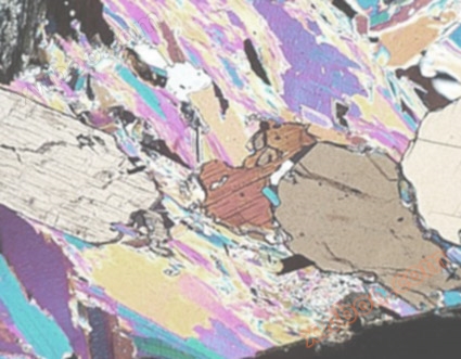


Example of testing precious medicinal herb ginseng:

|
1.resin canal |
Fragments are easily visible, and there are light yellow or yellow brown secretions in the cavity |
|
2.Clusters of calcium oxalate |
Large diameter, sharp or blunt edges, colorful under polarizing microscope |
|
3.Starch granules |
Single spherical, semi-circular or irregular polygonal, diameter4-20umThe umbilical point is dotted or fissured, Composite particles2classifying-6Particle composition, presenting a black cross shape under polarizing microscope |
|
4.catheter |
Mainly for mesh or ladder shaped conduits, with a diameter of10-56umThe pores of the mesh tube are relatively large |
|
5.cork cell |
On the surface, it appears to be square, rectangular or polygonal in shape, with thin walls and fine wavy bends. On the cross-section, the cells appear flat |
The necessity of polarizing mirror inspection:
1.Application in mineral based traditional Chinese medicine
The vast majority of mineral based traditional Chinese medicines are crystalline minerals, such as quartz, mica stone, and cold water stone. Strong and multi colored interference bands will appear in the dark field, which are very intuitive and obvious.
2.Application in animal based traditional Chinese medicine
The grinding plates of animal bones and teeth may not be visible under a regular microscope, but under a polarizing microscope, they exhibit strong color and stripe contrasts. Polarization can also distinguish normal cells from tumor cells, as normal cells often exhibit left-handed polarization, while tumor cells tend to exhibit right-handed polarization; Some animals' striated muscles and hair can also be examined using a polarizing microscope. Many animal medicinal herbs such as leopard bone, ivory, sea dragon, seahorse, turtle shell, whole armor, snakes, and even many animal horn, stone, secretion medicinal herbs such as deer antlers, antelope antlers, bezoar, horse treasure, monkey jujube, pearl, musk, bird's nest, etc. can be examined using a polarizing microscope.
3Application in plant-based medicinal materials
Many plant medicinal materials exhibit stable and specific polarization phenomena in their tissues, cells, and contents. Using polarizing glasses for observation can quickly and accurately identify identification features and eliminate interference
3.1Starch granules
All plant starch granules exhibit polarization phenomenon. Under a polarizing microscope, in a dark background, starch granules appear as bright spherical shapes with two intersecting transient stripes, and the intersection point is the navel of the starch granules.
TIPS: Microscopic specimen preparation
Microscopic identification of complete medicinal materialsSuitable for observing the complete tissue structure of medicinal materials. Firstly, select the appropriate parts of the medicinal herbs and make microscopic specimens before observing them under a microscope. When making films, most medicinal herbs can be sliced horizontally, and some even need to be sliced vertically. The slicing methods include manual slicing, sliding slicing, paraffin slicing, etc. Among them, the manual slicing method is the most simple, fast, and commonly used. Hand cut slices must be processed and sealed with appropriate solutions in order to clearly observe tissue structure and the shape of cells and their contents. Sometimes, in order to observe certain cellular tissues such as fibers, ducts, etc., it is also necessary to prepare tissue dissociation slices and powder slices. When observing the residual content in the tissue slices or powders of medicinal materials, glycerol acetic acid test solution or distilled water is generally used to observe the starch granules. Polarized microscopy can also be used to observe the polarization phenomenon of unglazed starch granules; Observe paste powder particles, starch particles, oil droplets, resin, etc. using dilute glycerol tablets; Using hydrated chloral solution, inulin can be immediately observed without heating. If heated, it can dissolve starch granules, proteins, chloroplasts, resins, volatile oils, etc., and cause contracted cells to expand, resulting in clear cell tissue and significant characteristics of calcium oxalate crystals.
Microscopic identification of broken medicinal materialsFor large pieces of medicinal herbs, tissue slices can also be cut, and the method is generally the same as the microscopic identification method for intact medicinal herbs; When the block is small and difficult to slice, chemical reagents can be used to dissect the plant tissue and make dissociated tissue slices for observation, or scrape them off
Microscopic identification of powdered medicinal materials and observation of powder slices. For chopped grass and leaf medicinal herbs, surface observation is still the most commonly used method.Processing and sealing methods of powder materials and microscopic identification of intact medicinal materials
be similar. The powder flakes made can undergo microscopic chemical reactions. Identification of authenticity and purity based on the characteristics of tissue fragments and post contents observed under a microscope. The microscopic identification method of traditional Chinese patent medicines and simple preparations, such as pills, powders and chongs, is basically the same as that of powder medicinal materials. Measuring the size of cells and their contents is also an important basis for microscopic identification. In particular, the size of the same kind of cells or after contents in traditional Chinese patent medicines and simple preparations can often be used as a distinguishing feature between varieties.Microscopic identification of mineral drugs