-
E-mail
sales@sgm7.com
- Phone
-
Address
No. 10 San Yue Road, Pujiang Town, Shanghai
Product Categories
Shanghai Huatong Optical Instrument Co., Ltd
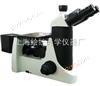
Inverted metallographic microscope 4XB-C | Metallographic analyzer price - Huatong Optical Factory
NegotiableUpdate on 01/11
- Model
- Nature of the Manufacturer
- Producers
- Product Category
- Place of Origin
Overview
Eyepiece: With anti mold function, flat field 10X high eye point eyepiece, field of view 22mm, high eye point observation, pupil distance 21mm, adjustable refractive power. *Objective: Infinite flat field achromatic objective (with anti mold function), 4X/0.10; 10X/0.25; 40X/0.65 (with spring and buffer device); 100X/1.25 (oil, with spring and buffer device).
Product Details
| Inverted metallographic microscope4XB-C ▲Inverted metallographic microscopeMainly used to identify and analyze the microstructure of various metals and alloys, applied in factories or laboratories for casting quality identification, raw material inspection, or research and analysis of metallographic structure after material treatment. ▲Can identify and analyze the microstructure of various metals, alloy materials, non-metallic substances, integrated circuits, micro particles, wires, etc ▲The 4XB-C inverted metallographic microscope has the advantages of multiple accessories, wide applicability, and the ability to perform bright field, dark field, and polarized observation. ▲Provide users with various image acquisition modes such as digital photography professional CCD, high-definition DV, etc,Not only can it observe dynamic images, but it can also edit, save, and print the required images And measurement and post-processing work | |
| 4XB-CInverted metallographic microscopeconfiguration table | |
| Component Name | Technical Specifications |
| magnification | Optical amplification: 100-500X; System amplification: 100-2000X |
| fuselage | Integrated structure, imported design scheme, overall die-casting of the body, stable and reliable structure |
| Infinite chromatic aberration independent correction optical system | |
| Equipped with a camera interface on the body | |
| Rotary color filter set (blue, green, yellow, 50% dimming film), built-in | |
| Focusing mechanism: Low position coaxial coarse and fine adjustment handwheel, fine adjustment handwheel grid value 0.001mm | |
| Wide voltage, 110∽240V,50/60Hz | |
| Observation tube | Three eye observation tube, tilt angle of 45, pupil distance adjustment of 55-75mm, adjustable visual acuity |
| eyepiece | WF10X/20mm, High Eyepoint |
| Metallographic objective lens | Infinite flat field achromatic objective 10X |
| Infinite flat field achromatic objective 20X | |
| Infinite flat field achromatic objective 50X | |
| Lighting lightbox | 12V50W/100W halogen light box |
| power distribution box | 12V/50W halogen lamp power box |
| Polarization component | Polarizing mirror plugin |
| Polarizer plugin | |
| Carrier platform | Mechanical moving stage 350mm × 208mm, moving range 50mm X50mm |
| Water droplet carrier (φ 118) | |
| Objective lens converter | 4. Internal positioning hole converter (suitable for balance field metallographic objective) |
| C-type interface | 0.5X camera adapter (C interface) |
| Lighting bulb | 12V50W Osram halogen bulb |
| fuse tube | 250V/5A Φ5X20 |
| Micrometer | 1/100mm |
| wrench | 2mm internal hexagonal wrench |
| 2.5mm internal hexagonal wrench | |
| 3mm internal hexagonal wrench | |
| power cord | 3C certified power cord |
| dust cover | dust cover |
| packaging | 4XB-C Instruction Manual |
| Foam upper cover, foam base, plastic bag (used for host packaging, environmental protection materials) | |
| System Composition | Computer type (4XB-C): 1invertmetallographic microscope2. Adaptation lens 3, high-definition camera 4, computer |
| Digital type (4XB-D)1invertmetallographic microscope2. Adaptation mirror 3, digital camera | |
. Scientific grade lossless format image output and storage
. High fidelity color reproduction using natural color matrix technology
. Global white balance and regional white balance functions
. Design of anti electromagnetic interference structure
. Convenient and fast one click device software installation, one click image acquisition and storage function
. Rich selection of photography interface accessories, suitable for the vast majoritymicroscope,Biological Microscope,polarizing microscopeMetallographic microscope,Phase contrast microscope, etc
| serial number | pixel | resolution | photography | category | layout | interface | connection method |
| 01 | 300ten thousand | 2048 X 1536 | 3.2MP | Color CMOS | 1/2” | C-Mount | USB2.0 |
| 02 | 520ten thousand | 2592 X 1944 | 5.2MP | Color CMOS | 1/2” | C-Mount | USB2.0 |
| 03 | 800ten thousand | 3264 X 2448 | 8.0MP | Color CMOS | 1/2” | C-Mount | USB2.0 |
| 04 | 910ten thousand | 3488 X 2616 | 9.1MP | Color CMOS | 1/2” | C-Mount | USB2.0 |
| 05 | 1000ten thousand | 3664 X 2748 | 10MP | Color CMOS | 1/2” | C-Mount | USB2.0 |
| 06 | 1400ten thousand | 4384 X 3288 | 14MP | Color CMOS | 1/2” | C-Mount | USB2.0 |
| The above cameras are all equipped with measurement, processing, segmentation, fusion and other functional software, with clear effects and easy use | |||||||
HToup View software Introduction to Image Processing Software
| Image acquisition: | The resolution size, capture and storage format, image attributes, color, brightness, contrast, exposure, white balance and other parameters of the image can be set, and can be used for photography, video recording, timed photography, etc. |
| Image measurement: | The image can be measured for parameters such as perimeter, perimeter, angle, area, circle diameter, and ellipse length and diameter, for example, using tools such as short lines, rectangles, irregular shapes, ellipses (circles), three-point circles, etc., and the parameters can be exported in EXCEL formatIt can synchronously display the image size ratio (i.e. scale bar), making the image size positioning more accurate |
| Image processing: | Real time dynamic adjustment of brightness, chromaticity, saturation, and red-green-The blue color can be adjusted to perform image processing such as color inversion, relief, sharpening, smoothing, graying, noise removal, rotation, flipping, and mirroring on the captured image. |
| Annotated drawing: | Convenient and efficient text annotation, arrow indication, and various geometric shape annotations. |
| Image segmentation: | Monochrome and multi-color binary threshold adjustment, corrosion and pore removal functions further accurately delineate contours, and segment images |
| Image stitching: | When the microscope can only capture partial images of the specimen, arrange the obtained partial images in order, and then use the image stitching function to obtain the global image of the entire specimen for research and storage. |
| Image Fusion: | When the thickness of the specimen is uneven or there is a height difference on the surface, due to the limitation of the depth of field of the high-power objective lens, only clear local images can be observed. Therefore, images from different focal planes can be captured, and the image fusion function can be used to obtain a complete and clear image. |
| quantity statistics | Quickly count the quantity and provide parameters such as overall and individual perimeter and area, which can be used for overall or local statistics, and the measured data can be exported. |
| Graphic and textual report: | Help you easily create experimental reports that combine text and images, provide detailed textual explanations for specimen images, and print them. |
Each set of cameras includes:
| digital camera (Standard)CInterface1.8riceUSBCable) | 1 |
| TCN-0.5Photography receiver,23,30,30.5Millimeter direct insertion interface (optional) | 1 |
| 0.01Millimeter micrometer (optional) | 1 |
| Driver and software CD | 1 |
| Certificate of Conformity | 1 |
| carton packaging | 1 |
| Product advantages: |
| Sony Japan(SONY)CompanyExView HADseriesCCDimage sensor |
| The image processing circuitUltra-FineTMColor engine rendering technology |
| sensor chipPCBACenter pre calibration process (error ±)2Pixel) |
| comply withISO9001TheISO14001TheRoHSTheCETheFCCWaiting for quality and environmental certification standards |
| |
| Product Features: |
| Extremely high sensitivity |
| Extremely high quantum efficiency |
| Extremely high signal-to-noise ratio |
| Extremely low dark current |
| Extremely low readout noise |
| Ultra long controllable exposure time |
| Advanced white balance settings |
| ROIVariable area |
| 1X1The2X2The4X4Pixel overlay enhancement(BINNING) |
| Fully enclosed high-quality aluminum alloy metal shell |
| Support platform drivers and applications:Microsoft WindowsTheMacTheLinux |
| Support multiple videosAPIinterfaceDirectShowTheTwainTheLabView |
| Data output interface:USB2.0TheUSB3.0 |
| Optical interface:C-MountTheCS-MountTheF-Mount |
| |
| Application fields: |
| |
| Bright field, phase contrast, and dark field microscopes |
| |
| Pathology, histology, and cytology |
| |
| Immunofluorescence microscope |
| |
| Green fluorescent protein(GFP)The application |
| |
| fluorescence in situ hybridization |
| |
| Calcium ion ratio analysis |
| |
| Vitality and Movement Analysis |
| |
| Deoxyribonucleic acid analysis |
| |
| metallographic microscope |
| |
| semiconductor inspection |
| |
| Manufacturing Quality Control |
| |
| Failure Analysis |
| |
| Forensic analysis |
| |
| Hematoxylin-Eosin staining observation (pathology) |
| |
| Gel imaging |
| |
| semiconductor inspection |
| |
| Solar panel inspection |
http://www.htxwj.com/ Industrial Microscope
http://www.htxwj.com/ microscope
http://www.htxwj.com/ Biological Microscope
http://www.htxwj.com metallographic microscope
http://www.htxwj.com polarizing microscope
http://www.htxwj.com Mineralogical microscope
http://www.htxwj.com Phase contrast microscope
http://www.omshtong.com/ Industrial Microscope
http://www.omshtong.com/ microscope
http://www.omshtong.com/ Biological Microscope
http://www.omshtong.com metallographic microscope
http://www.omshtong.com polarizing microscope
http://www.omshtong.com Mineralogical microscope
http://www.omshtong.com Phase contrast microscope
Similar Product Recommend


