-
E-mail
smkj526@163.com
-
Phone
18100753880吴,18175146520邹
-
Address
Building S5, Chuanghui Business Center (AUX Center), No. 182 Xiaoxiang South Road, Yuelu District, Changsha City, Hunan Province
Product Categories
Hunan Shengming Technology Co., Ltd
Olympus Confocal Microscope OLS5000
NegotiableUpdate on 03/16
- Model
- Nature of the Manufacturer
- Producers
- Product Category
- Place of Origin
Overview
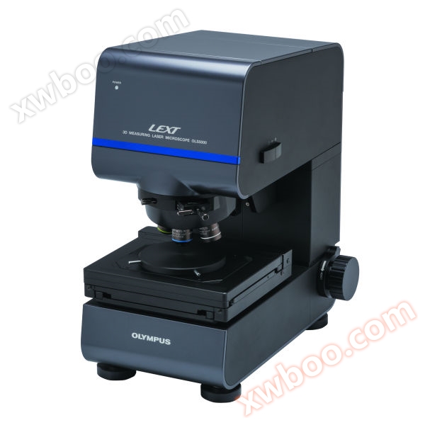
Olympus 3D Laser Confocal Microscope LEXTOLS50003D Measurement Laser Confocal Microscope adopts advanced optical system, which can generate high-quality images through non-destructive observation for accurate 3D measurement, ISO standard surface roughness measurement, etc. Its preparation work is also very simple and does not require pre-processing of the sample
Product Details
Olympus 3D Laser Confocal Microscope LEXT OLS5000
The Olympus OLS5000 3D measurement laser confocal microscope adopts an advanced optical system, which can generate high-quality images through non-destructive observation for accurate 3D measurement, ISO standard surface roughness measurement, etc. Its preparation work is also very simple and does not require pre-processing of the sample.
LEXT OLS5000 3DThe two optical systems (color imaging optical system and laser confocal optical system) equipped on the measuring laser microscope enable it to obtain color information, height information, and high-resolution images.
LEXT OLS5000 3DMeasurement laser microscope has4Key Value:
·Capture any surface shape.
·Quickly obtain reliable data.
·Easy to use-Just place the sample and press the button once.
·Measure challenging samples.
value1Capture any surface shape.
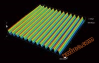
OLS5000The advanced technology of microscopes enables them to perform high-resolution imaging3DSample measurement.
value2Quickly obtain reliable data
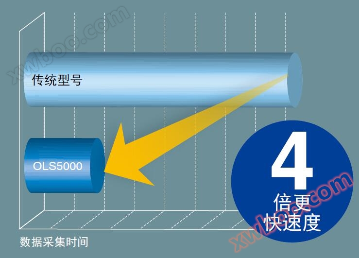
The scanning algorithm of this microscope can improve both data quality and speed, thereby shortening your scanning time and simplifying your workflow,zuiUltimately achieving an improvement in productivity.
value3Easy to use, just place the sample and press the button once
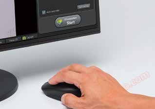
LEXT ® OLS5000The microscope has an automatic data acquisition function, so there is no need to make complex settings adjustments. Even unfamiliar users can obtain accurate detection results.
value4Measurable and challenging samples
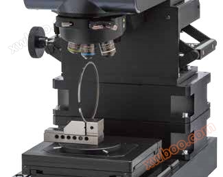
Low output, non-contact non-destructive laser measurement means no sample preparation is required. It is possible to measure vulnerable materials without damaging them. Expansion rack can accommodate up to210Millimeter samples, while ultra long working distance objectives can measure depths up to25Millimeter dents. When measuring these two types of samples, you only need to place the samples on the stage.
[Obtain color information]
The use of white light in color imaging optical systemsLEDLight source andCMOSThe camera captures color information.
[obtain3DHeight information and high-resolution confocal images]
The laser confocal optical system adopts405Confocal images were obtained using a nano laser diode light source and a high-sensitivity photomultiplier tube. Shallow focal depth enables it to be used for measuring surface irregularities of samples.
[405Nano laser light source]
The lateral resolution of optical microscopes improves as the wavelength decreases. Compared to using visible light (peak value), laser microscopes using short wavelength lasers550Traditional microscopes with nanometer resolution have better lateral resolution.OLS5000Microscope utilization405Nano short wavelength laser diodes achieve excellent lateral resolution.
[Laser confocal optical system]
The laser confocal optical system only receives light focused through a circular pinhole, and does not collect all light reflected and scattered from the sample. This helps to eliminate blurring, allowing it to obtain images with higher contrast than ordinary microscopes
[X-Yscanner]
OLS5000The microscope is equipped with an Olympus optical scanner. By utilizing electromagnetic inductionMEMSThe resonant scannerXAxis and adoptionGalvanoScanning galvanometerYCombining axes can enableX-YThe scanner is positioned at a conjugate position relative to the objective lens and pupil, thus achieving excellent results with low scanning trajectory distortion and minimal optical aberrationX-YScan.
[Principle of Height Measurement]
When measuring height, the microscope obtains multiple confocal images by automatically moving the focal position.
Based on discontinuous focal positions(Z)And detecting light intensity(I)It is possible to estimate the intensity variation curve of each pixel(I-ZObtain the peak position and peak intensity of the curve. Due to the peak positions of all pixels corresponding to the irregularity of the sample surface, it is possible to obtain the surface of the sample3DShape information. Similarly, peak intensity data can be used to obtain images (extended images) of focal points at all positions on the sample surface.
Host specifications:
|
model |
OLS5000-SAF |
OLS5000-SMF |
OLS5000-LAF |
OLS5000-EAF |
OLS5000-EMF |
||
|
Total magnification |
54x - 17,280x |
||||||
|
Field of view diameter |
16 μm - 5,120 μm |
||||||
|
measuring principle |
optical system |
Reflective confocal laser scanning laser microscope |
|||||
|
Light receiving element |
Laser: Photomultiplier tube (2ch) |
||||||
|
Height measurement |
display resolution |
0.5 nanometre |
|||||
|
dynamic range |
16 position |
||||||
|
Repeatability n-1 * 1 * 2 * 6 |
20x : 0.03μm, 50x : 0.012 μm, 100x : 0.012 μm |
||||||
|
Accuracy * 1 * 3 * 6 |
Measurement value+/-1.5% |
||||||
|
Splicing image accuracy * 1 * 4 * 6 |
20x : 15+0.5Lμ m, 50x: 9+0.5L μ m, 100x: 7+0.5L μ m (L: splicing length [μ m]) |
||||||
|
Measurement noise (SQ noise) * 1 * 5 * 6 |
1 nanometre |
||||||
|
width measure |
display resolution |
1 nanometre |
|||||
|
Repeatability 3 n-1 * 1 * 6 |
20x : 0.05μm, 50x : 0.04 μm, 100x : 0.02 μm |
||||||
|
Accuracy * 1 * 3 * 6 |
Measurement value+/-1.5% |
||||||
|
Splicing image accuracy * 1 * 3 * 6 |
20x : 15+0.5Lμ m, 50x: 9+0.5L μ m, 100x: 7+0.5L μ m (L: splicing length [μ m]) |
||||||
|
The largest number of measurement points in a single measurement |
4096 x 4096pixel |
||||||
|
The largest number of measurement points |
3600 megapixel |
||||||
|
XYStage configuration |
Length measurement module |
• |
not have |
not have |
• |
not have |
|
|
scope of work |
100 x 100 mmelectric |
100 x 100 mmhand movement |
300 x 300 mmelectric |
100 x 100 mmelectric |
100 x 100 mmhand movement |
||
|
zuiLarge sample height |
100 mm |
30 mm |
30 mm |
210 mm |
140 mm |
||
|
laser light source |
wavelength |
405 nm |
|
||||
|
zuilarge output |
0.95 mW |
|
|||||
|
Laser classification |
2 Class (IEC60825-1:2007, IEC60825-1:2014) |
|
|||||
|
Colored Lighting |
White LED |
||||||
|
Electrical power |
240 W |
240 W |
278 W |
240 W |
240 W |
||
|
quality |
Microscope body |
About 31 kilograms |
About 30 kilograms |
About 50 kilograms |
About 43 kilograms |
About 39 kilograms |
|
|
controller |
About 12 kilograms |
||||||
| |
|||||||